Home » Without Label » Anatomy Of Ribs : Structure Of The Ribcage And Ribs : You will also find the xiphoid process, 10th rib, the apex of the heart, the coronary vein of the heart.
Anatomy Of Ribs : Structure Of The Ribcage And Ribs : You will also find the xiphoid process, 10th rib, the apex of the heart, the coronary vein of the heart.
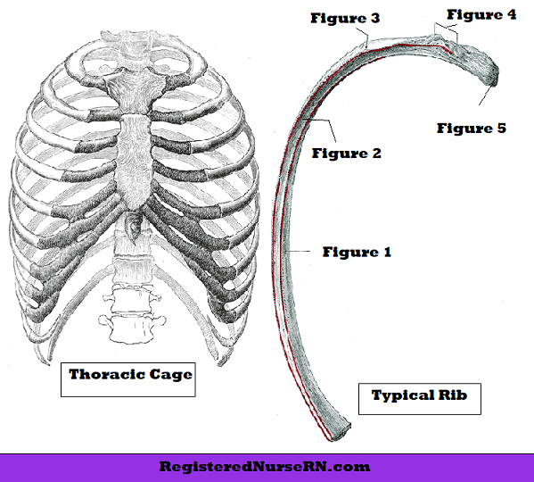
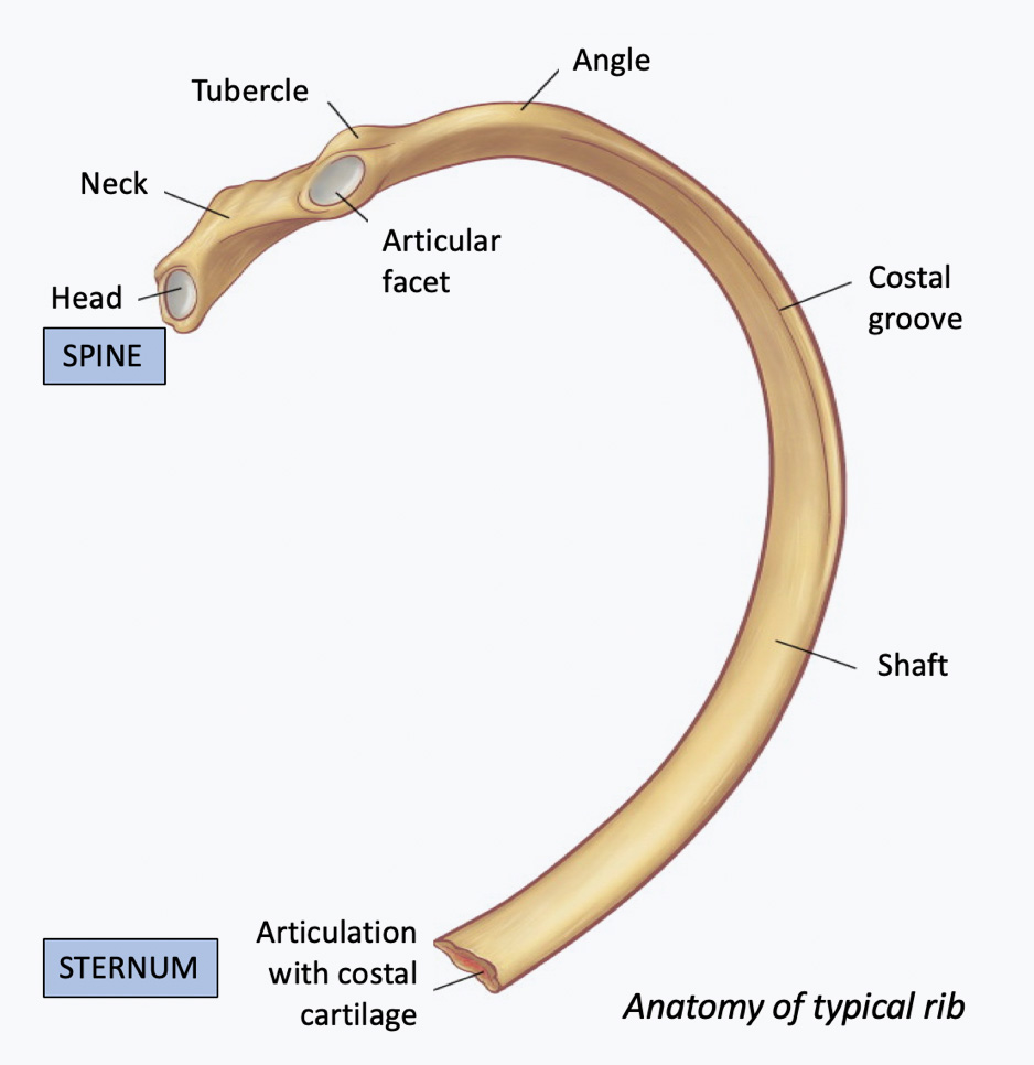
Anatomy Of Ribs : Structure Of The Ribcage And Ribs : You will also find the xiphoid process, 10th rib, the apex of the heart, the coronary vein of the heart.. As part of the bony thorax, the ribs protect the internal thoracic organs. It functions in movement, respiration, and protection of the thoracic. In this video, you will learn the bony features of typical and atypical ribs. It consists of the 12 pairs of ribs with their costal cartilages and the sternum (figure 6.38). Costae) are long, flat, curved bones that form the rib cage.
Your heart sits in the middle of your chest, to the left. The first seven ribs progressively increase in length, the lower five ribs then begin to decrease in length. Lumbar ribs have been reported to be more numerous than cervical ribs (8% of individuals). There are twelve pairs of ribs, all of which articulate with the vertebral column. Of these, 35% were cervical ribs.
Rib Bone Anatomy Quiz from www.registerednursern.com Ribs the ribs partially enclose and protect the chest cavity, where many vital organs (including the heart and the lungs) are located. Anatomy the rib cage is a bony structure found in the chest (thoracic cavity). Contributing to their role in protecting the internal thoracic organs. The 1 st, 11 th and 12 th ribs are considered atypical ribs due to. 16 photos of the rib cage diagram with organs. The joint of head of rib (costocorporeal joint) is the articulation between the rib head and vertebral body. 12 photos of the anatomy of ribs and its related area. The first seven true ribs connect to the sternum via the costal cartilages by day 45.
Anatomy the rib cage is a bony structure found in the chest (thoracic cavity).
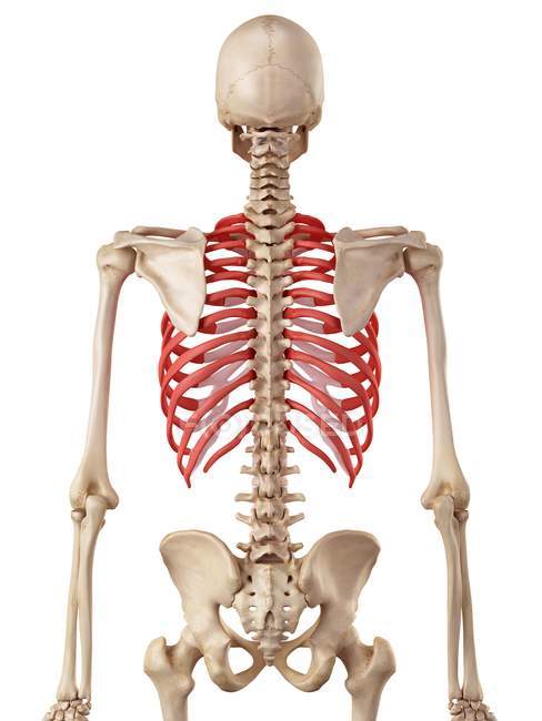
Ribs are highly vascular and trabecular with a thin outer layer of compact bone. Extra levator costae muscles may be associated with these ribs. As part of the bony thorax, the ribs protect the internal thoracic organs. It functions in movement, respiration, and protection of the thoracic. Lumbar ribs are usually quite small and seldom as complete as cervical ribs. 12 photos of the anatomy of ribs and its related area. They are extremely light, but highly resilient; The flexible (hyaline) cartilage, makes the breathing process easier. Secondary ossification centers appear at 15 years (, 1,, 2). The ribs are a set of 12 pairs bones which form the protective 'cage' of the thorax. The thoracic wall is formed by 12 ribs, 12 thoracic vertebrae, cartilage, sternum, and five muscles. The rib cage surrounds the lungs and the heart, serving as an important means of bony protection for these vital organs.in total, the rib cage consists of the 12 thoracic vertebrae and the 24 ribs, in addition to the sternum. The 1 st, 11 th and 12 th ribs are considered atypical ribs due to.
The 4 main types of pork ribs explained. Contributing to their role in protecting the internal thoracic organs. The heart is a muscle at the center of your circulatory system. The flexible (hyaline) cartilage, makes the breathing process easier. Anatomy the rib cage is a bony structure found in the chest (thoracic cavity).
Rib Injury Rib Injury Clinic from www.ribinjuryclinic.com We can't talk about the different cuts of pork ribs without looking at the anatomy of the pig. Extra levator costae muscles may be associated with these ribs. The anatomy of the human ribs (costae) are one of the integral parts of the chest wall; The main functions of this glandular organ are the secretion of enzymes for the digestion of fat, and maintenance. The bones of the rib cage are the sternum, the 12 thoracic vertebrae and the 12 pairs of ribs. The ribs are a set of 12 pairs bones which form the protective 'cage' of the thorax. Ribs anatomy the average skeleton contains 24 individual ribs, formed in 12 pairs, and they are divided into three main categories: Anatomy the rib cage is a bony structure found in the chest (thoracic cavity).
Each pig has 14 rib bones which are attached to the spine and divided into the four most popular cuts.
In this image, you will find common carotid arteries, internal jugular vein, subclavian artery, subclavian vein, heart, right lung, 6th rib, diaphragm, costal cartilage in it. One of the internal organs on right side of body is the liver, which is also the largest glandular organ. Rib development and normal anatomy twelve paired ribs develop from cartilaginous costal processes of the developing thoracic vertebrae. Your heart sits in the middle of your chest, to the left. 12 photos of the anatomy of ribs and its related area. The sternum is a flat bone that is made up of three parts, the (1) manubrium, (2) body, and the (3) xiphoid process. The first seven sets of ribs, known as true ribs also known as vertebrosternal ribs, are directly articulate with the vertebral column posteriorly and terminate anteriorly as costal cartilage. As part of the bony thorax, the ribs protect the internal thoracic organs. Each pig has 14 rib bones which are attached to the spine and divided into the four most popular cuts. True ribs, false ribs, and floating ribs. They are extremely light, but highly resilient; Choose from 500 different sets of anatomy of the ribs flashcards on quizlet. The ribs are curved, flat bones which form the majority of the thoracic cage.
Anatomy, types, ossification & clinical significance. Chest bone, ribs, lung, heart, xiphoid process, sternum anatomy. In this video, you will learn the bony features of typical and atypical ribs. There are 12 pairs of ribs which are separated by intercostal spaces. One of the internal organs on right side of body is the liver, which is also the largest glandular organ.
Human Rib Cage Anatomy Human Physiology Osteology Stock Photo 160568256 from st.focusedcollection.com Each pig has 14 rib bones which are attached to the spine and divided into the four most popular cuts. Chest bone, ribs, lung, heart, xiphoid process, sternum anatomy. 16 photos of the rib cage diagram with organs. The joint of head of rib (costocorporeal joint) is the articulation between the rib head and vertebral body. It functions in movement, respiration, and protection of the thoracic. According to medical news today, it's important to understand the anatomy of the rib cage when determining whether pain under the rib cage is mild enough for home treatment, or severe enough to seek medical attention. There are 12 pairs of ribs which are separated by intercostal spaces. Anatomy the rib cage is a bony structure found in the chest (thoracic cavity).
The ribs are curved, flat bones which form the majority of the thoracic cage.
Anatomy the rib cage is a bony structure found in the chest (thoracic cavity). Each pair is numbered based on their attachment to the sternum, a bony process at the front of the rib cage which serves as an anchor point. A thorough comprehension of the anatomy and function of the thorax will help identify, differentiate, and treat the plethora of pathology that can occur within the thorax. Extra levator costae muscles may be associated with these ribs. 12 photos of the anatomy of ribs and its related area. 16 photos of the rib cage diagram with organs. The top edge of the manubrium has a depression called the suprasternal or jugular notch. You will also find the xiphoid process, 10th rib, the apex of the heart, the coronary vein of the heart. The rib cage surrounds the lungs and the heart, serving as an important means of bony protection for these vital organs.in total, the rib cage consists of the 12 thoracic vertebrae and the 24 ribs, in addition to the sternum. In this article, we review rib development and normal anatomy and techniques for evaluating the ribs and present inherited abnormalities and acquired conditions of the pediatric ribs. The 1 st, 11 th and 12 th ribs are considered atypical ribs due to. In this image, you will find common carotid arteries, internal jugular vein, subclavian artery, subclavian vein, heart, right lung, 6th rib, diaphragm, costal cartilage in it. The ribs are a set of twelve paired bones which form the protective 'cage' of the thorax.